Home
/ Histology Of Smooth Muscle Diagram - Histology Of Muscle Tissue - Vsmcs display diversity in function and phenotype depending on their location within the arterial tree (large conduit vs.
Histology Of Smooth Muscle Diagram - Histology Of Muscle Tissue - Vsmcs display diversity in function and phenotype depending on their location within the arterial tree (large conduit vs.
Histology Of Smooth Muscle Diagram - Histology Of Muscle Tissue - Vsmcs display diversity in function and phenotype depending on their location within the arterial tree (large conduit vs.. This page describes smooth muscle development, descriptions of cardiac muscle and smooth muscle development can be found in other notes. I am sherif ibrahim a lecturer of histology and cell biology and graduate of michigan state university, usa. The contaction of smooth muscle cells is involuntary and the neuromuscular junctions controlling. Kierszenbaum, al histology and cell biology 2nd ed., mosby elsevier, 2007, p. Exception :smooth muscle of iris originate from ectoderm.
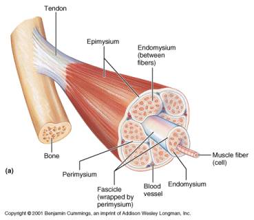
When you swallow, tension is applied to. The diagram is fully labeled. The position of smooth muscle within the wall of the intestine is illustrated by light microscopy in figure a. In a motor unit the motor neuron branches to form neuromuscular. Smooth muscle (trachea histological slide).
Histology Lab 6 Muscles Flashcards Quizlet from o.quizlet.com Smooth muscle also lines the majority of the digestive system, for similar reasons. The contaction of smooth muscle cells is involuntary and the neuromuscular junctions controlling. Here, the smooth muscle fibers are organized into two. The histology online learning module has clear and concise aims, objectives and anticipated outcomes, listed below. The diagram is fully labeled. It is divided into two subgroups; The nuclei of smooth muscle fibers are euchromatic, centrally located and oval shaped. In this thin section, use the same criteria as described above for webslide #32 to distinguish between.
The smooth muscle in the intestine is arranged into two layers:
These will appear on the. An inner circular layer and an outer longitudinal layer. The nuclei of smooth muscle fibers are euchromatic, centrally located and oval shaped. Here is a cross section of the ileum. There are 3 different types of muscle: The arrangement of smooth muscle differs from organ to organ. From wikimedia commons, the free media repository. Kierszenbaum, al histology and cell biology 2nd ed., mosby elsevier, 2007, p. The position of smooth muscle within the wall of the intestine is illustrated by light microscopy in figure a. It is composed of thin actin g. They produce connective tissue proteins such as collagen and elastin for which reason they are also referred to as fixed (or stationary). However, the cells in the digestive system have different stimuli than those in the circulatory system. Microscopic view of smooth muscle.
Histology Of Muscle from faculty.etsu.edu Each muscle fibre may contain numerous longitudinal fibrils called. One important histologic feature is the smooth muscle component of. I am sherif ibrahim a lecturer of histology and cell biology and graduate of michigan state university, usa. Here is a cross section of the ileum. This page describes smooth muscle development, descriptions of cardiac muscle and smooth muscle development can be found in other notes. Kierszenbaum, al histology and cell biology 2nd ed., mosby elsevier, 2007, p. Differences between smooth cardiac and skeletal muscle. This section of dentaljuce has over 400 histological slides, showing tissues from all organ systems in their healthy state.
Download scientific diagram | histology and smooth muscle actin (sma) the research progress in anatomy and histology of the complex of levator palpebrae superioris and müller's muscle.
In a motor unit the motor neuron branches to form neuromuscular. Human anatomy for muscle, reproductive, and skeleton. I am sherif ibrahim a lecturer of histology and cell biology and graduate of michigan state university, usa. The muscle fibres contains specialized cytoplasm called sarcoplasm the muscle fibres may be bounded by the cell membrane called sarcolemma. Myofibroblasts represent a special type of smooth muscle cell which additionally have qualities of fibrocytes. Smooth muscle occurs in snug parallel bundles, with more nuclei and with nuclei all internal to fibers. This page describes smooth muscle development, descriptions of cardiac muscle and smooth muscle development can be found in other notes. An inner circular layer and an outer longitudinal layer. There are 3 different types of muscle: The histology online learning module has clear and concise aims, objectives and anticipated outcomes, listed below. In this thin section, use the same criteria as described above for webslide #32 to distinguish between. The contaction of smooth muscle cells is involuntary and the neuromuscular junctions controlling. Kierszenbaum, al histology and cell biology 2nd ed., mosby elsevier, 2007, p.
Related posts of smooth muscle diagram labeled. In a motor unit the motor neuron branches to form neuromuscular. In this thin section, use the same criteria as described above for webslide #32 to distinguish between. This section of dentaljuce has over 400 histological slides, showing tissues from all organ systems in their healthy state. From wikimedia commons, the free media repository.